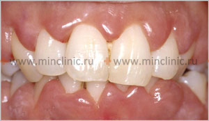
Chronic hypertrophic gingivitis
Understanding Chronic Hypertrophic (Hyperplastic) Gingivitis
Definition and Etiology
Chronic hypertrophic gingivitis, also referred to as hyperplastic gingivitis or gingival enlargement, is a chronic inflammatory condition of the gingival (gum) tissue characterized by an increase in its size or volume. This overgrowth can be localized to specific areas or generalized throughout the mouth. While local factors like dental plaque are often involved in initiating or exacerbating the inflammation, systemic factors frequently play a significant role in the development and nature of the hypertrophy.
The primary basis for the occurrence of many forms of chronic hypertrophic gingivitis involves changes in the patient's systemic status, particularly:
- Hormonal Fluctuations: Changes in hormonal status are a major contributing factor. This is seen in:
- Endocrine diseases (e.g., thyroid disorders, though less commonly a direct cause of true hypertrophy).
- Puberty (pubertal gingivitis).
- Pregnancy (pregnancy gingivitis, pregnancy "tumor" or pyogenic granuloma).
- Menopause.
- Use of hormonal contraceptives.
- Drug-Induced Gingival Overgrowth: Certain medications are well-known to cause gingival enlargement as a side effect. These include:
- Anticonvulsants (e.g., phenytoin/Dilantin).
- Calcium channel blockers (e.g., nifedipine, amlodipine, verapamil) used for hypertension and cardiovascular conditions.
- Immunosuppressants (e.g., cyclosporine A) used in organ transplant recipients or for autoimmune diseases.
- Systemic Diseases: Conditions like leukemic reticulosis (a form of leukemia) or other hematological disorders can manifest with gingival enlargement and inflammation.
- Chronic Irritation and Inflammation: Poor oral hygiene leading to chronic plaque accumulation can cause inflammatory gingival enlargement, which is a common type. Mouth breathing can also contribute to anterior gingival inflammation and hypertrophy.
- Genetic/Hereditary Factors: Hereditary gingival fibromatosis is a rare genetic condition causing generalized fibrous gingival enlargement.
- Chronic Intoxication: Exposure to certain toxins (less common).
Clinical Manifestations and Key Features
Chronic hypertrophic gingivitis is primarily manifested by an increase in the volume of the gingival papillae (the gum tissue between teeth) and/or the marginal gingiva (the edge of the gum surrounding the teeth). This enlargement can lead to the formation of false periodontal pockets (pseudopockets). These are deepened gingival sulci created by the overgrown gingival tissue, not by the destruction of periodontal attachment and bone loss as seen in true periodontal pockets (periodontitis).
Key characteristics of chronic hypertrophic gingivitis include:
- Preservation of Periodontal Attachment: The epithelial gingival attachment to the tooth remains at its normal level (at or near the cementoenamel junction). There is no apical migration of the junctional epithelium.
- Absence of Alveolar Bone Loss: There are no pathological changes or loss in the bone tissue of the alveoli (the bony sockets supporting the teeth) directly attributable to the gingivitis itself.
- The severity of enlargement can range from mild (involving only papillae) to moderate (involving papillae and marginal gingiva) to severe (covering a significant portion of the tooth crowns).
Forms of Chronic Hypertrophic Gingivitis
Based on clinical and morphological (histopathological) changes, two main forms of chronic hypertrophic gingivitis are distinguished: the edematous form and the fibrous form.
Edematous Form: Morphology and Clinical Picture
Morphologically, the edematous form of hypertrophic gingivitis is primarily characterized by:
- Significant edema (swelling due to fluid accumulation) of the connective tissue elements within the gingival papillae and marginal gingiva.
- Vasodilation (enlargement of blood vessels) and increased vascular permeability.
- Swelling and degeneration of collagen fibers.
- A prominent inflammatory infiltrate, often rich in lymphocytes and plasma cells (lymphoplasmacytic tissue infiltration).
The clinical picture of the edematous form of hypertrophic gingivitis typically includes patient complaints of:
- An aesthetic defect due to the unusual, swollen appearance of the gums.
- Pain or soreness in the gums, especially when brushing teeth or during eating.
- Easy gingival bleeding, often spontaneous or with slight provocation.
Upon examination of the oral cavity:
- The gingival papillae and marginal gingiva are visibly enlarged, soft, boggy, and edematous.
- The color is often bright red (hyperemic) or bluish-red (cyanotic) due to vascular engorgement and stasis.
- The gums bleed easily upon gentle probing or manipulation.
- The surface of the papillae often has a smooth, glossy, or shiny appearance due to the edema.
- A characteristic sign is that after pressing on the surface of the swollen papilla with the blunt part of a dental instrument (e.g., a periodontal probe), a temporary depression or pitting may remain.
- Varying amounts of dental deposits (plaque and calculus) may be found, which contribute to the inflammation.
Fibrous Form: Morphology and Clinical Picture
Morphologically, the fibrous form of hypertrophic gingivitis is primarily characterized by:
- Proliferation (overgrowth) of dense fibrous connective tissue elements within the gingival papillae and marginal gingiva.
- Coarsening and increased density of collagen fibers.
- Epithelial changes such as acanthosis (thickening of the spinous layer) and parakeratosis (retention of nuclei in the stratum corneum).
- Edema and inflammatory tissue infiltration are typically not prominent or are much less expressed compared to the edematous form. The tissue is more cellular and less vascular.
The clinical picture of the fibrous form of hypertrophic gingivitis often involves patient complaints primarily focused on:
- The unusual, enlarged appearance of the gums and the associated aesthetic defect.
- Pain is usually absent or minimal.
- Bleeding is typically not a significant feature, or occurs only with more vigorous trauma.
Upon examination of a patient with the fibrous form of hypertrophic gingivitis:
- The gingival papillae and marginal gingiva are determined to be enlarged, firm, dense, and resilient to the touch.
- The color is often pale pink, similar to healthy gingiva, or slightly pinker.
- Soreness on palpation and bleeding on probing are usually absent or minimal.
- The surface may appear lobulated or nodular.
- Hard and soft subgingival dental deposits can often be found, as the enlarged tissue can create deeper pseudopockets that are difficult for the patient to clean effectively.
It is also possible for mixed forms to exist, exhibiting features of both edematous and fibrous hypertrophy.
Diagnosis of Hypertrophic Gingivitis
The diagnosis of hypertrophic gingivitis is usually straightforward for a dental professional and is based on clinical findings. To assess the patient's dental status and confirm the diagnosis, the following are typically sufficient:
- Patient History (Questioning): Including medical history (medications, systemic conditions, hormonal status like puberty or pregnancy), dental history, onset and nature of gingival changes, and oral hygiene habits.
- Clinical Examination: Visual inspection and palpation of the gums to assess size, shape, color, consistency, surface texture, and extent of enlargement.
- Probing of Clinical Pockets (Pseudopockets): To measure the depth of the gingival sulcus/pseudopocket and assess for bleeding on probing. It is crucial to differentiate pseudopockets from true periodontal pockets (which involve attachment loss).
- Schiller-Pisarev Test (Historical/Adjunctive): An iodine-based staining test that highlights areas of gingival inflammation due to increased glycogen content; more likely to be positive and intense in the edematous form.
In doubtful cases, especially if the enlargement is localized, rapidly growing, or has an unusual appearance, an X-ray examination (e.g., periapical or panoramic radiographs) is indicated to rule out underlying bony pathology or involvement characteristic of periodontitis (which would show bone loss). In hypertrophic gingivitis, alveolar bone levels should be normal.
To exclude systemic conditions like blood diseases (e.g., leukemia, which can cause gingival enlargement), all patients with significant or atypical hypertrophic gingivitis should undergo a general blood test (Complete Blood Count - CBC with differential). Patients should also be consulted by or referred to specialist doctors of the appropriate profile if systemic etiological factors are suspected (e.g., gynecologist for pregnancy-related changes, endocrinologist for hormonal imbalances, hematologist if blood dyscrasias are suspected). In some cases, an in-depth study of the patient's hormonal status (e.g., hormone assays) may be required.
A biopsy of the enlarged gingival tissue may be necessary in some cases, particularly if:
- The diagnosis is uncertain.
- The lesion is localized and atypical.
- Malignancy needs to be ruled out.
- To confirm drug-induced gingival overgrowth or hereditary gingival fibromatosis.
Treatment of Chronic Hypertrophic Gingivitis
General Principles
The treatment of chronic hypertrophic gingivitis is carried out taking into account the identified etiological factors, the specific morphological picture (edematous or fibrous), and the clinical form and severity of the disease. The primary goals are to reduce gingival inflammation and enlargement, restore normal gingival contour, and establish and maintain good oral hygiene to prevent recurrence.
Initial therapy for all forms typically includes:
- Identification and Elimination of Etiological Factors:
- Meticulous professional removal of all dental plaque and calculus (scaling and root planing).
- Correction of any plaque-retentive factors (e.g., overhanging restorations, poorly fitting appliances).
- Intensive oral hygiene instruction and motivation for the patient.
- If drug-induced, consultation with the patient's physician to consider alternative medications if possible.
- Management of underlying systemic conditions or hormonal imbalances in collaboration with medical specialists.
Treatment of the Edematous Form of Hypertrophic Gingivitis
In the edematous form of hypertrophic gingivitis, treatment initially focuses on controlling the acute inflammation and edema:
- Anti-inflammatory Therapy:
- Thorough removal of dental deposits as described above.
- Topical applications of anti-inflammatory and antimicrobial agents (e.g., chlorhexidine rinses or gels, metronidazole gel).
- Prescription of anti-inflammatory physiotherapy modalities such as galvanization, iontophoresis (e.g., with corticosteroids or NSAIDs), or darsonvalization (high-frequency electrotherapy).
- Sclerotherapy (for persistent edema unresponsive to initial therapy): This involves the application or injection of sclerosing (hardening or scarring) agents to reduce tissue volume by inducing fibrosis.
- Topical Application: Imposing on the gingival margin and introducing into the clinical pseudopockets tampons or pellets moistened with various sclerosing compounds, such as:
- 20-30% resorcinol solution (historical).
- 10-25% zinc chloride solution (historical).
- 5-10% alcoholic propolis solution.
- Injections (Deep Sclerosing Therapy): If topical application sclerotherapy is ineffective, injections of hypertonic solutions directly into the gingival papillae may be performed under local anesthesia. Examples include:
- 10% calcium chloride solution.
- 40-60% glucose solution.
- 10% calcium gluconate solution.
- 90% ethyl alcohol solution (used with extreme caution due to necrotizing potential).
- Topical Application: Imposing on the gingival margin and introducing into the clinical pseudopockets tampons or pellets moistened with various sclerosing compounds, such as:
- Topical Corticosteroids: Steroid hormones can be used as decongestants for the edematous form. This may involve injections into the papillae of 0.1-0.2 ml of hydrocortisone emulsion, or daily rubbing into the gingival papillae of ointments containing glucocorticoid hormones (e.g., Ftorocort, Lorinden, Deperzolone, Hyoxysone - brand names may vary). Glucocorticoids can also be incorporated into gingival dressings (periodontal packs). Use of potent corticosteroids should be short-term and monitored due to potential side effects.
- Home Care: Patients with the edematous form are prescribed rinses and mouth baths with herbal decoctions (e.g., chamomile, sage, calendula) for their mild anti-inflammatory properties, alongside meticulous oral hygiene.
- Surgical Excision (Gingivectomy): If conservative treatment (including sclerotherapy) for the edematous form of hypertrophic gingivitis proves ineffective and significant enlargement persists, surgical excision of the hypertrophied gingival margin – a gingivectomy operation – is performed. This is typically done under local anesthesia, often addressing a segment of 6-8 teeth simultaneously. The excision of the hypertrophied gums is performed with an incision that starts coronally to the base of the pseudopocket (closer to the transitional fold/mucogingival junction if very enlarged) and goes obliquely towards the tooth surface at the bottom of the "false" pocket. This excises the outer part of the hypertrophied gingival margin, recontouring the gingiva to a more physiological shape.
Treatment of the Fibrous Form of Hypertrophic Gingivitis
The fibrous form of hypertrophic gingivitis, being characterized by dense connective tissue proliferation, is less responsive to anti-inflammatory and sclerosing therapies. Management often involves:
- Cytotoxic Drugs (Historical/Experimental Use): The use of cytotoxic drugs, for example, novembichin (an alkylating agent), has been historically described for fibrous forms. A solution (e.g., 10 mg of the drug dissolved in 10 ml of isotonic sodium chloride solution) was injected into the hypertrophied papillae (0.1-0.2 ml weekly, for a course of 3-5 injections). This approach is highly specialized, carries significant risks, and is not a standard or widely accepted treatment in modern periodontology due to toxicity concerns.
- Diathermocoagulation (Electrosurgery): Point diathermocoagulation of hypertrophied gingival papillae can be effective for the fibrous form. The operation is performed under local anesthesia. An electrode (e.g., a root canal needle or specialized electrosurgical tip) is inserted into the papilla tissue to a depth of 3-5 mm. In one session, 4-5 papillae might be coagulated. This technique ablates excess tissue.
- Surgical Excision (Gingivectomy/Gingivoplasty): Most often, the fibrous form of chronic hypertrophic gingivitis requires surgical excision of the overgrown gingival tissue. A gingivectomy (excision of gingiva to eliminate pseudopockets and recontour the tissue) or gingivoplasty (reshaping of the gingiva) is performed to restore normal gingival architecture and facilitate oral hygiene.
Special Considerations: Pregnancy, Juvenile Gingivitis, Leukemia
- Pregnancy Gingivitis (often hypertrophic, edematous type): In pregnant patients with hypertrophic gingivitis, initial treatment focuses on meticulous removal of dental deposits and intensive anti-inflammatory therapy (professional cleaning, oral hygiene instruction, chlorhexidine rinses). Surgical interventions are generally deferred until after childbirth if possible. If the condition of the gums does not return to normal postpartum, then sclerotherapy or surgical methods may be considered.
- Juvenile (Adolescent/Pubertal) Hypertrophic Gingivitis: Often related to hormonal changes and sometimes exacerbated by orthodontic appliances or suboptimal hygiene. A more conservative, wait-and-see approach is often taken initially, with primary efforts focused on maintaining excellent oral hygiene and professional debridement. Treatment of chronic hypertrophic gingivitis is typically performed if the pathological changes in the gums do not resolve spontaneously after the end of puberty.
- Leukemia-Associated Gingival Enlargement: In patients with leukemia, dentists carry out only symptomatic therapy for the chronic hypertrophic gingivitis component (e.g., gentle debridement, chlorhexidine rinses to control secondary infection and discomfort). Sclerosing agents, most physiotherapeutic modalities, and elective surgical methods of treatment are generally contraindicated in this situation due to bleeding risks, impaired healing, and the need to manage the underlying systemic disease first. Periodontal management is done in close consultation with the patient's hematologist/oncologist.
Differential Diagnosis of Gingival Enlargement
Gingival enlargement can be caused by various factors, and a correct diagnosis is crucial:
| Condition | Key Differentiating Features |
|---|---|
| Chronic Hypertrophic Gingivitis (Inflammatory) | Edematous or fibrous enlargement, often related to plaque and local irritants, may be exacerbated by hormones. No true attachment loss. |
| Drug-Induced Gingival Overgrowth | History of taking specific medications (phenytoin, cyclosporine, calcium channel blockers). Enlargement often fibrous, starts in papillae, can become generalized. May or may not have significant inflammation if hygiene is good. (May relate to hypertrophic gingivitis if inflammatory component is strong). |
| Hereditary Gingival Fibromatosis | Genetic predisposition; firm, dense, pale pink generalized gingival enlargement, often starting with tooth eruption. Not primarily inflammatory. (Can be considered a type of periodontoma or idiopathic condition). |
| Gingivitis/Periodontitis Associated with Systemic Conditions (e.g., Leukemia, Vitamin C deficiency/Scurvy) | Leukemia: often boggy, hemorrhagic, sometimes necrotic enlargement. Scurvy: swollen, bleeding, purplish gums. Requires systemic diagnosis and management. (See also Periodontitis for general periodontal inflammation). |
| False Enlargement (due to underlying bony exostoses or tori) | Gingiva appears enlarged due to prominent underlying bone, not true gingival tissue increase. Bone is hard on palpation. |
| Benign or Malignant Gingival Tumors (e.g., Epulis, Squamous Cell Carcinoma) | Often localized, may have different texture/color, rapid growth, ulceration, or induration if malignant. Biopsy essential. (Related to Periodontomas). |
Complications and Prevention
Untreated chronic hypertrophic gingivitis can lead to:
- Aesthetic concerns.
- Difficulty with oral hygiene due to altered gingival contours and pseudopockets, leading to increased plaque accumulation and risk of caries or progression to periodontitis.
- Pain and discomfort, especially with the edematous form.
- Speech or mastication difficulties if enlargement is severe.
Prevention and management involve:
- Meticulous personal oral hygiene.
- Regular professional dental cleanings.
- Addressing underlying systemic factors or drug regimens where possible.
- Early intervention if gingival changes are noted.
When to Seek Dental Care
Individuals should consult a dentist or periodontist if they notice:
- Persistent swelling or enlargement of their gums.
- Gums that bleed easily, are red, or tender.
- Changes in the appearance or contour of their gums.
- Difficulty cleaning around enlarged gum tissue.
- If they are taking medications known to cause gingival overgrowth.
Early diagnosis and management can prevent further progression and improve outcomes.
References
- Newman MG, Takei HH, Klokkevold PR, Carranza FA. Carranza's Clinical Periodontology. 13th ed. Elsevier; 2019. (Covers gingival enlargement extensively)
- Hassell TM. Phenytoin-induced gingival overgrowth. In: Myers HM, ed. Monographs in Oral Science. Vol. 9. Karger; 1981.
- Trackman PC, Kantarci A. Molecular and clinical aspects of drug-induced gingival overgrowth. J Dent Res. 2015 Apr;94(4):540-6.
- Mariotti A. Dental plaque-induced gingival diseases. Clin Periodontol. 2004;5(1):7-19.
- American Academy of Periodontology. Task Force Report on the Update to the 1999 Classification of Periodontal Diseases and Conditions. J Periodontol. 2015 Jul;86(7):835-8. (Context for classifying gingival diseases)
- Chapple ILC, Mealey BL, Van Dyke TE, et al. Periodontal health and gingival diseases and conditions on an intact and a reduced periodontium: Consensus report of workgroup 1 of the 2017 World Workshop on the Classification of Periodontal and Peri-Implant Diseases and Conditions. J Periodontol. 2018 Jun;89 Suppl 1:S74-S84.
- Rateitschak KH, Rateitschak EM, Wolf HF, Hassell TM. Color Atlas of Dental Medicine: Periodontology. 3rd ed. Thieme; 2005.
See also
- Dental anatomy
- Dental caries
- Periodontal disease:
- Chronic catarrhal gingivitis
- Chronic generalized periodontitis of moderate severity
- Chronic hypertrophic gingivitis
- Chronic mild generalized periodontitis
- Idiopathic periodontal disease, periodontomas
- Periodontitis
- Periodontitis in remission
- Periodontosis
- Severe chronic generalized periodontitis
- Ulcerative gingivitis


