Seeing what kinds of tissues form your body
Organizing cells into tissues
A tissue is an assemblage of cells, not necessarily identical but from the same origin, that together carry out a specific function. As we discuss in Chapter 1, tissue is the second level of organization in organisms, above (larger than) the cell level and below (smaller than) the organ level.
Like just about everything else in anatomy, tissues are many and various, and they’re grouped into a reasonable number of "types" to make talking about them and understanding them a little simpler. The tissues of the animal body are grouped into four types: connective tissue, epithelial tissue, muscle tissue, and nervous tissue. All body tissues are classified into one of these groups.
Connecting with connective tissue
Connective tissues connect, support, and bind body structures together and are the most abundant tissue by weight. Generally, connective tissue is made up of cells that are spaced far apart within a gel-like, semisolid, solid, or fluid matrix. (A matrix is a material that surrounds and supports cells. In a chocolate chip cookie, the dough is the matrix for the chocolate chips.)
Connective tissue has many functions, and thus many forms; it is the most varied of all the tissue groupings. In some parts of the body, such as the bones, connective tissue supports the weight of other structures, which may or may not be directly connected to it. Other connective tissue, like adipose tissue (fat pads), cushions other structures from impact. You encounter lots of connective tissue in the chapters to come because every organ system has some kind of connective tissue.
We discuss the specialized connective tissues bone and cartilage in some detail in Chapter 5, and we discuss the important connective tissue blood in detail in Chapter 9. (What? Blood is a tissue? A connective tissue? Yes, and you’ll see why.)
The other types of connective tissue, or proper connective tissues, are all means of connections. Just as we have different kinds of tapes and glues, we have different varieties of connective tissue. They contain varying proportions of fibrous proteins of two types: collagenous and elastic. Collagenous fibers are made of collagen, a bulky protein, and serve to provide structure. Elastic fibers are made of elastin, a thin protein, and serve to provide stretch.
The following are the proper connective tissues found in the human body:
- Areolar (a type of loose connective tissue): This tissue surrounds and separates structures in every part of the body. It forms a thin membrane with ample space for blood vessels to pass through. Both collagenous and elastic fibers are prevalent in its gel-like matrix.
- Dense regular connective tissue: This tissue is characterized by its tightly packed collagenous fibers arranged parallel to each other, making it very strong. There are very few cells present and there is very little blood flow. Tendons and ligaments are made of dense regular tissue (see Chapter 5).
- Dense irregular connective tissue: Very similar to dense regular, the fibers in this tissue are disorganized, leaving more space for blood flow. The dermis is a typical tissue of this type (see Chapter 4).
- Adipose tissue (a type of loose connective tissue): Composed of fat cells, adipose tissue provides fuel storage and insulation as well as support and protection to its underlying structures.
- Reticular tissue (a type of loose connective tissue): This type of tissue uses a thinner variety of collagenous fibers to form a net. It creates the framework of such organs as the spleen, the lymph nodes, and the liver.
- Elastic connective tissue: Collagenous fibers are bundled in parallel with bands of elastic fibers sandwiched between (like lasagna). This makes elastic tissue strong but stretchy — perfectly suited for the walls of hollow organs and arteries.
Continuing with epithelial tissue
Epithelial tissue forms the epidermis of the integument (the skin and its accessory structures; see Chapter 4), covers all your internal organs, and forms the lining of the internal surfaces of the blood vessels and hollow organs.
Epithelial tissues create coverings and linings; they’re always bordered by "empty space" on one side. The other side is a basement membrane that allows resources to diffuse up into the tissue from the connective tissue below (epithelial tissues have no blood flow).
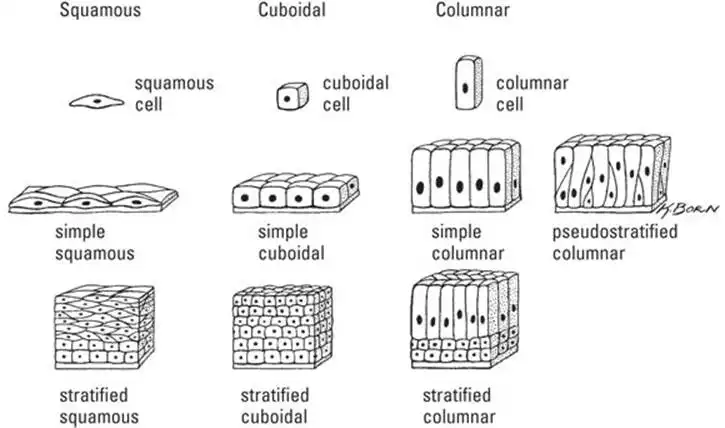
Epithelial tissue comes in eight types that are defined by the way epithelial cells are combined and shaped (see Figure 3-8).
- Simple squamous epithelium: A single layer of flat cells, this tissue functions in rapid diffusion and filtration. The lining of the alveoli (small sacs) of the lung is a typical tissue of this type.
- Simple cuboidal epithelium: A single layer of cuboidal cells, this tissue functions in absorption and secretion. This tissue is typically found in glands. The cuboidal cells have the capacity to produce and modify the glandular product (for example, sweat, oil, or milk).
- Simple columnar epithelium: A single layer of cells that are elongated in one dimension (like a column). Like simple cuboidal epithelium, this tissue functions in secretion and absorption. This type of tissue is primarily found lining portions of the digestive tract.
The cells may also be ciliated, possessing a type of organelle called cilia — hairlike structures that act to move substances along in waves. Ciliated simple columnar epithelium can be found lining the uterine tube. - Pseudostratified columnar epithelium: A single layer of columnar cells. Note that the prefix pseudo means "false." The tissue appears stratified, or layered, because the cells’ nuclei don’t line up in a row, as they do in simple columnar epithelium. Other than that, they’re the same, and have similar functions of absorption and secretion. This type of tissue lines ducts in testicular structures.
More commonly, this tissue type is ciliated. It is present in the linings of the respiratory tract, functioning in a more or less identical way to the simple columnar ciliated epithelium. - Stratified squamous epithelium: This tissue consists of several layers of cells: squamous epithelial cells on the outside with deeper layers of cuboidal or columnar epithelial cells. It’s found in areas where the outer layer is subject to wear and needs to be replaced continuously. The epidermis of the skin is an example of a specific type called keratinized stratified squamous epithelial tissue.
- Stratified cubiodal epithelium: Several layers of cuboidal cells that line the ducts associated with the sweat, mammary, and salivary glands.
- Stratified columnar epithelium: Several layers of elongated cells mainly act as protection in such structures as the conjunctiva of the eye and the pharynx.
- Transitional epithelium: The cells of this tissue can change (or transition) from cuboidal when relaxed to squamous when stretched, as needed by the tissue. This tissue is found in the lining of the bladder, where having a little room to stretch is sometimes handy. See a diagram of transitional epithelium in Chapter 12.
Mixing it up with muscle tissue
Muscle tissue comes in three types: skeletal muscle, smooth muscle, and cardiac muscle. We discuss the similarities and differences in cellular composition in these three types of tissue in Chapter 6. We also discuss in Chapter 6 the anatomy and physiology of the large organ system called the muscular system, of which skeletal muscle tissue is a major component. In Chapter 9, we discuss the function of cardiac muscle in the context of the cardiovascular system as well as the role of smooth muscle in blood circulation. We also cover smooth muscle’s role in the digestive system in Chapter 11.
Getting nervous about nervous tissue?
Don’t be. Nervous tissue is relatively simple in one way: Your body has only one type of nervous tissue, and it’s made mostly of only one type of cell, the neuron. You can get lots more information about the nervous system in Chapter 7, if you get the impulse to find out more.
See also
- Locating Physiology on the Web of Knowledge
- Chapter 1. Anatomy and Physiology: The Big Picture
- Chapter 2. What Your Body Does All Day
- Chapter 3. A Bit about Cell Biology
- Sizing Up the Structural Layers
- Chapter 4. Getting the Skinny on Skin, Hair, and Nails
- Chapter 5. Scrutinizing the Skeletal System
- Chapter 6. Muscles: Setting You in Motion
- Talking to Yourself
- Chapter 7. The Nervous System: Your Body’s Circuit Board
- Chapter 8. The Endocrine System: Releasing Chemical Messages
- Exploring the Inner Workings of the Body
- Chapter 9. The Cardiovascular System: Getting Your Blood Pumping
- Chapter 10. The Respiratory System: Breathing Life into Your Body
- Chapter 11. The Digestive System: Beginning the Breakdown
- Chapter 12. The Urinary System: Cleaning Up the Act
- Chapter 13. The Lymphatic System: Living in a Microbe Jungle
- Life’s Rich Pageant: Reproduction and Development
- Chapter 14. The Reproductive System
- Chapter 15. Change and Development over the Life Span
- The Part of Tens
- Chapter 16. Ten (Or So) Chemistry Concepts Related to Anatomy and Physiology
- Chapter 17. Ten Phabulous Physiology Phacts
- Supplemental Images

